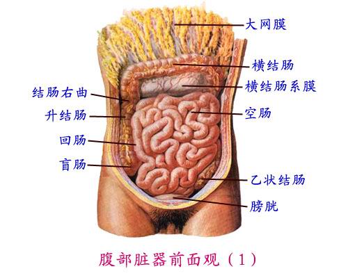
五、小肠 Fifth, the small intestine
(一)形态位置 (A) Form position
小肠长5~7米,位于腹腔中下部。 5 to 7 meters long small intestine, located in the lower part of the abdominal cavity. 上接幽门下续盲肠。 Continued on the next lower pyloric cecum.
(二)分部 (B) Division
小肠全长分十二指肠、空肠、回肠。 Total length of the small intestine points duodenum, jejunum, ileum.

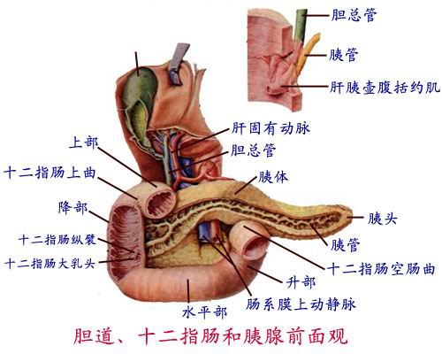
1. 1. 十二指肠 Duodenum
(1)上部:起自幽门,指向肝门。 (1) Upper: starting from the pylorus, point hilar. 起始处称十二指肠球,粘膜平滑,是溃疡好发部位。 Said at the start of duodenal mucosa smooth, ulcers predilection sites.
(2)降部:沿脊柱右侧下降,后内侧壁有十二指肠纵襞,末端为十二指肠大乳头,是胆总管和胰管的共同开口。 (2) descending part: the right side along the spine fall within the vertical rear wall duodenal folds, the end of the duodenal papilla, the common bile duct and pancreatic duct joint opening.
(3)水平部:向左横过第二腰椎。 (3) horizontal part: left across the second lumbar vertebra.
(4)升部:末端向下弯曲形成十二指肠空肠曲,被十二指肠悬肌(韧带)固定于腹后壁。 (4) l Department: duodenum, jejunum end bent downward song, was suspended muscle duodenum (ligament) is fixed to the abdominal wall. 十二指肠悬肌是上、下消化道的分界,也是临床外科手术识别空肠起端的标志。 Duodenum muscle is hanging on the boundaries of the lower digestive tract, but also the recognition of clinical surgery jejunum mark the borders.
2. 2. 空肠和回肠 Jejunum and ileum
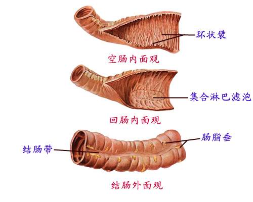
(1)空肠:腹腔左上部,空回肠前2/5,管径较大,管壁较厚,血供丰富。 (1) jejunum: abdominal cavity at the top left, front 2/5 ileum, large diameter, thick wall, blood supply.
(2)回肠:腹腔右下部,空回肠后3/5,管径略小,管壁较薄,血供稍差。 (2) ileum: celiac bottom right, after ileum 3/5, slightly smaller diameter, thin wall, somewhat less blood supply.
六、大肠 Six, large intestine
(一)形态位置 (A) Form position
大肠起自回肠,终于肛门。 Since the large intestine and ileum, and finally the anus. 约1.5米,呈“门框”状包绕在小肠周围。 About 1.5 meters, was "door" shape wrapped around the small intestine. 分为盲肠、结肠、直肠三部。 Divided into cecum, colon, rectum three. 盲肠和结肠表面有三个特征性的结构,即:结肠带、结肠袋、肠脂垂。 Cecum and colon surface have three characteristic structures, namely: the colon, colon bags, intestinal fat hanging. 结肠带共三条,交汇于阑尾根部。 Colon with a total of three, the intersection at the root of the appendix.
1. 1. 盲肠 Appendix
左接回肠,上续结肠,下连阑尾。 Left with ileum, colon on continued, even under appendix.
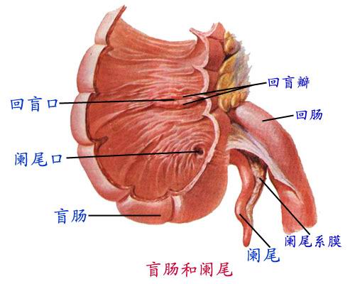
阑尾:长6~8cm,末端位置不定。 Appendix: Long 6 ~ 8cm, end position uncertain. 根部固定,恰是三条结肠带交汇处,是临床手术寻找阑尾的重要标志。 Roots fixed, precisely three colon with Interchange, is looking for clinical surgery appendix important symbol. 其体表投影为麦氏点,即脐与右髂前上棘连线中外1/3交点。 Its surface projection of Michael's point, that the navel and the front right iliac spine and foreign connections 1/3 intersection.
2. 2. 结肠 Colon
分为升结肠、横结肠、降结肠和乙状结肠。 Into the ascending colon, transverse colon, descending colon and sigmoid colon.
3. 3. 直肠 Rectum
上接乙状结肠,下接肛门,约15cm。 Continued sigmoid colon, then under the anus, about 15cm. 全长有两个弯曲,骶曲凸向后,会阴曲凸向前。 There are two full-length curved, convex curved backward sacral perineal convex curved forward. 以盆膈为界分为盆部和肛门部。 Pots bounded into pelvic diaphragm and anal.
(1)盆部:下份膨大,称直肠壶腹,内有2~3个直肠横襞,结肠镜检时应避免损伤。 (1) pelvic: In parts of enlargement, said the rectal ampulla, there are two to three horizontal folds rectum, colonoscopy should avoid damage.
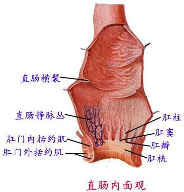
(2)肛管:主要结构有肛柱、肛瓣、肛窦、齿状线等。 (2) the anal canal: The main structure of the anal columns, anal lobe, anal sinus, dentate lines. 齿状线由肛柱下端和肛瓣边缘围成,线上线下的神经来源、动脉供应、血液回流均不一样,线上是粘膜,线下是皮肤。 Dentate line anal column by the lower edge of the flap and anal surrounded, online and offline sources nerves, arteries supply blood return are not the same, the line is a mucous membrane, the next line is the skin. 线上形成痔是内痔,线下形成痔是外痔,内痔不痛外痔痛。 The formation of hemorrhoids are hemorrhoids line, the line is forming hemorrhoids external hemorrhoids, internal hemorrhoids are not painful hemorrhoids pain.
齿状线构成、区别、意义 Dentate line constitutes the difference, meaning
肛柱下端肛瓣缘 Anal anal flap edge lower end of the column
连成锯齿环形线 Even a jagged ring line
线上粘膜下皮肤 Under the skin, mucous membranes online
供血回血分两端 Blood back to the ends of Blood
线下神经属躯体 The line somatic nerve genus
线上粘膜内脏管 Online mucosal gut tube
内痔不痛外痔痛 Internal hemorrhoids are not painful hemorrhoids pain
辛辣不吃酒莫沾 Do not stick spicy Chijiu