 +
+  +
+  +
+  =
= 
 +
+  +
+  +
+  =
= 
The fourth chapter
respiratory system
The first section respiratory tract
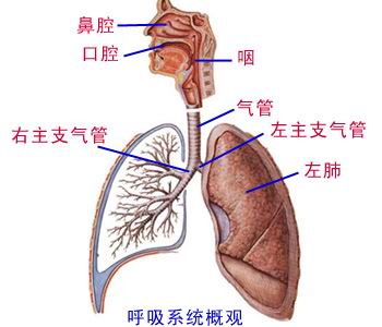
The respiratory tract including the nose, swallows, the throat, the trachea, the main bronchial tube. Under clinical general the nose, swallows, the throat to be called the upper respiratory tract, the trachea, the host bronchial tube is called the respiratory tract.
First, nose
(one) outside nose
Outside the nose assumes the sparganium stoloniferum cone-shape, take the bone and the cartilage as the support, outside becomes by the skin. The primary structure includes: Nose back, nose root, tip of the nose, wing of the nose, nostril and so on.
(two) nasal cavity
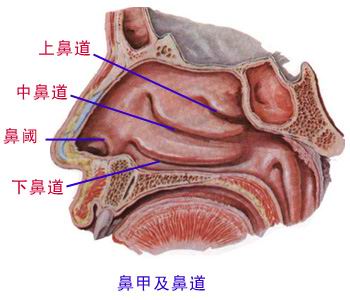
The nasal cavity is take the bone and the cartilage as the support, the inside lining mucous membrane and the skin becomes. The first hole passes the outside before the nose, after latter borrows the nose, Kong Tongyan. About middle is separated by the nose in divides into two. Each side divides the nose vestibule and the inherent nasal cavity. Outside the inherent nasal cavity on the sidewall has the upper, middle and lower three nose armor, underneath three nose armor respectively be upper, middle and lower three nasal passages. Before the next nasal passage, the share has the ductus nasolacrimalis aperture. The nasal cavity mucous membrane district is:
1. smells the area
Upside distributes separates the mucous membrane on nose armor and the nose, slightly assumes the buff color.
2. regio respiratoria:
Smells outside the area the mucous membrane, assumes the light red. Before and is located in the nose to separate, the lower part mucous membrane, the blood vessel is rich, said that easy to bleed the area.
Nasal cavity mucous membrane
On nose armor, on diaphragm Smells the area mucous membrane slightly light yellow
The regio respiratoria, occupies big The mucous edema is not unobstructed
Before the diaphragm, under easy to bleed The attention protection not damages
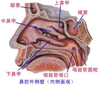
(three) paranasal sinus
The paranasal sinus constitutes by the osseous paranasal sinus inside lining mucous membrane.
1. upper jaw Dou
Located at the maxilla, opens the mouth in the nasal passage.
2. frontal sinus
Located at the frontal bone, opens the mouth in the nasal passage.
3. sinus ethmoidalis
Before, during and after located at the ethmoid bone, divides three crowds, first, group aperture in the nasal passage, latter group aperture on nasal passage.
4. sinus sphenoidalis
Located at the sphenoid bone, opens the mouth in the recessus spheno-ethmoidalis.
Paranasal sinus aperture spot
The tear-duct opens the mouth in most under Nasal mucus tear
Middle course frontal sinus upper jaw Dou Before the sinus ethmoidalis, not throws down
After the sinus ethmoidalis, on group nasal passage Recessus spheno-ethmoidalis only then it
Second, throat
(one) position
The jugal center the neck front part, 5~6 cervical vertebra altitudes, on Lian Shegu, gets down continues the trachea.
(two) structure
The throat is take the cartilage as a support, attaches by the throat muscle, the inside lining mucous membrane becomes.
1. laryngeal cartilage
Mainly has the thyroid cartilage, the cricoid cartilage, the arytenoid cartilage, the epiglottal cartilage. The thyroid cartilage taking advantage of armor shape tongue periosteum Lian Yushe the bone, gets down by the cricoid cartilage contracts the organization membrane taking advantage of the knot continually in the cartilago trachealis. Between laryngeal cartilage shape Cheng Huan armor, link scoop two groups of joints.
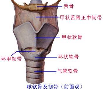
2. throat muscle
Two groups, affect Yu Huanjia, the link scoop joint separately, a group may intense or relaxes the vocal cord, a group expands or the reduction fissure of glottis.
Throat's structure
Armor shape ring-like scoop epiglottis Cartilage support ligament company
Surrounds the armor link scoop to be double-hinged Two group of throat myo- functions entire
Expanded reduction fissure of glottis The vocal cord loose and tight it also pulls
3. laryngeal cavity
(1) laryngeal cavity structure
1) vestibule bi: Two bi are the vestibule cracks.
2) sound bi: Two bi are the fissures of glottis. Is the narrowest spot.
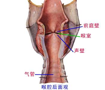
(2) branch
1) vestibulum laryngis: Is located at vestibule crack above laryngeal cavity, with swallows after the throat opening is interlinked, is spacious.
2) throat middle cavity: Is located at between the vestibule crack and the fissure of glottis the laryngeal cavity, is the laryngeal cavity three narrowest spots.
3) under glottis cavity: Located at fissure of glottis below. Under its mucous membrane the organization is loose, when inflammation easy to have dropsy to create the acute laryngemphraxis.
Third, trachea
(one) position
Trachea before the neck center, front esophagus. Continues in the sixth cervical vertebra body lower limb plane meets Yu Hou the cricoid cartilage.
(two) constitutes
The trachea is “C” the body and spirit tube cartilage link contracts the organization and the smooth muscle by 16~20 taking advantage of the knot links becomes.
(three) branch
The trachea cuts the mark take breastbone's neck vein as to divide into the pate and the chest.
1. trachea pate
front 2~4 cartilago trachealis link has the thyroid gland canyon, the clinical tracheotomy often chooses 3~4 or 4~5 cartilago trachealis link place.
2. chest
In the angulus sterni plane branch is the left and right main bronchial tube.
Trachea constitution, branch
C body and spirit tube cartilage link Six neck lower limbs continue Yu Huan
About chest angle plane minute The entire journey is divided the neck chest section
Neck Duan Qianyou thyroid gland The incision needs to elect five to three
Fourth, main bronchial tube
(one) morphological property
Left host bronchial tube tall and slender, near level. The right host bronchial tube stubby, nearly is vertical.
(two) significance
The trachea foreign matter crashes enters the right main bronchial tube.
About main bronchial tube characteristic, significance
The bronchial tube, divides nearby two Left tall and slender right stubby